(RxWiki News) Once cancer is diagnosed, imaging tests are usually performed to see if the cancer has spread. Medical imaging specialists (radiologists) usually perform these tests, but that shouldn’t always be the case, according to a new review.
A team of researchers suggested that the surgeon should perform ultrasound tests of thyroid cancer patients prior to surgery rather than relying on ultrasounds performed by radiologists.
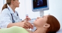
Ultrasound uses sound waves to produce images.
The recent analysis revealed that ultrasounds performed by surgeons prior to surgery discovered nearly a third more diseased lymph nodes that needed to be removed during surgery.
"Ask questions about any medical test ordered for you."
Kevin Parrack, MD, endocrine surgery instructor at the New York-Presbyterian Hospital/Columbia University Medical Center, New York, NY, and colleagues reviewed more than 12 years of thyroid cancer cases.
The researchers were looking at the impact of ultrasounds performed by a surgeon in assessing and executing the correct surgical approach for patients with thyroid cancer that had spread to the lymph nodes. Lymph nodes are found throughout the body and play a key role in the immune system.
The thyroid is shaped like a butterfly and wraps around the windpipe. This organ secretes hormones that influence the metabolism. Some 60,000 cases of thyroid cancer will be diagnosed in Americans this year.
The cases reviewed involved patients with differentiated thyroid cancer, which has cancer cells that look very similar to normal cells.
Thyroid ultrasounds are generally performed by radiologists. But these tests don’t typically evaluate lymph nodes near the thyroid.
The authors of this review pointed out that a clinician cannot feel diseased lymph nodes, so relying only on radiologist reports can lead to surgeries that don’t remove all of the cancer.
The researchers reviewed the cases of 137 individuals who had undergone surgery to treat differentiated thyroid cancer between 2000 and 2013 at their referral center.
Of these patients, 94 had imaging of the neck prior to being referred to a surgeon. Imaging tests included ultrasounds, CT (computed tomography) scans and MRI (magnetic resonance imaging).
A surgeon performed the initial ultrasound for the remaining 6 percent of the patients.
All of the patients then had another ultrasound by an endocrine surgeon prior to surgery.
Surgeon performed ultrasounds detected and confirmed diseased lymph nodes in 31 percent of the patients who’d received imaging tests by a radiologist.
A total of 55 patients had undergone ultrasounds performed by a radiologist before being referred to a surgeon. Of these patients, 35 percent had diseased lymph nodes that the surgeon's ultrasound picked up. Detecting these lymph nodes changed the surgical plan significantly.
Julie Ann Sosa, MD, professor of surgery and medicine at Duke Clinical Research Institute in Durham, NC, said in a prepared statement, “These data are significant in that they suggest the surgeon is uniquely positioned to perform ultrasound in a way that it affords critical information that would not otherwise be available for optimizing surgical approach."
Findings from this study were presented at 83rd Annual Meeting of the American Thyroid Association.
All research is considered preliminary before being published in a peer reviewed journal.