(RxWiki News) Parkinson’s disease can be hard for doctors to identify. And while it has no cure, early diagnosis can help those with the nerve-damaging disease choose an optimum course of treatment.
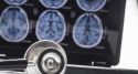
Ultra-high-field magnetic resonance imaging (MRI) gave a detailed look at the part of the brain affected by Parkinson’s, according to a new study.
The authors of this study concluded that this high-powered MRI may lead to earlier diagnosis.
"See your doctor if you notice tremors or shaking in a limb."
Mirco Cosottini, MD, from the University of Pisa in Italy, was lead author of this study.
From April 2012 through April 2013, Dr. Cosottini and his fellow researchers analyzed the brains of 38 people. Of that group, 17 had Parkinson's disease and 21 did not. The Parkinson’s patients ranged in age from 38 to 70. Those without Parkinson’s ranged from 25 to 62 years old.
To determine the accuracy of this type of MRI in detecting Parkinson’s, these researchers also compared those 38 brains to a single, non-Parkinson’s brain of a woman who had died of a heart attack at age 67.
Using the ultra-high-field 7-Tesla (7-T) MRI, these researchers were able to distinctly view the three layers of a quarter-moon shaped group of cells in the mid-brain named substantia nigra (SN). When cells in the SN that produce dopamine die, the brain begins to lose its ability to control such things as mood, stress, addiction and motor skills. The body naturally makes dopamine, a chemical that stimulates the nervous system and controls various bodily functions.
The lack of motor skills can result in an extreme degree of bodily trembling, stiffness, lack of balance and coordination for Parkinson’s patients.
After these researchers finished doing the 7-T MRIs, they trained two neuroradiologists in how to read these MRIs.
Based on the neuroradiologists’s readings of mid-brain abnormalities in the Parkinson’s patients, Dr. Cosottini and his team determined that the ultra-high-field 7-T MRI let the neuroradiologists correctly classify all (100 percent) of the Parkinson’s patients as having that disease. The neuroradiologists also correctly classified 96.2 percent of the healthy participants as not having Parkinson’s.
Traditional MRIs are less powerful than the 7-T and typically have not been used to diagnose Parkinson’s because their ability to do so is limited, Dr. Cosottini and team wrote. Instead, physicians have relied on a person’s medical history and a neurological exam that involves measuring such functions as hearing, speech, vision, mental wellness, coordination and other physical skills and reflexes controlled by the nervous system.
It is often difficult to distinguish Parkinson's disease from other conditions using those methods alone, these researchers added.
“ … [T]he possibility of reaching a diagnostic result by using a short [242-second MRI sequence] … is an important goal of 7-T MR imaging. Because at present 7-T MR imaging systems are authorized exclusively for research purpose and not for clinical use, the demonstration of their clinical usefulness and their safety will be the conditions required for their acceptance in the medical community."
According to Dr. Cosottini, results from this study could pave the way for earlier detection of the disease through the extra powerful MRI and, as a result, earlier treatment.
"Parkinson's disease diagnosis remains clinically based, but with the introduction of 7-T MRI into clinical practice, a supporting radiologic diagnosis can be made," he said.
This study was published March 5 in Radiology.
The study report did not list any funders for this research.
This team of researchers included three who've received research grants to install MRI systems.